Keratoconus in Teens and Tweens
I'm sure there are tons of articles and posts out there about keratoconus. But, in the past month, I have diagnosed 3 kids with keratoconus - two of whom were previously misdiagnosed by another eye doctor, so I thought it might be good to get something out there focusing on this disease in kids.First, what is keratoconus? Let's go back to our diagram of the eyeball I drew a while ago.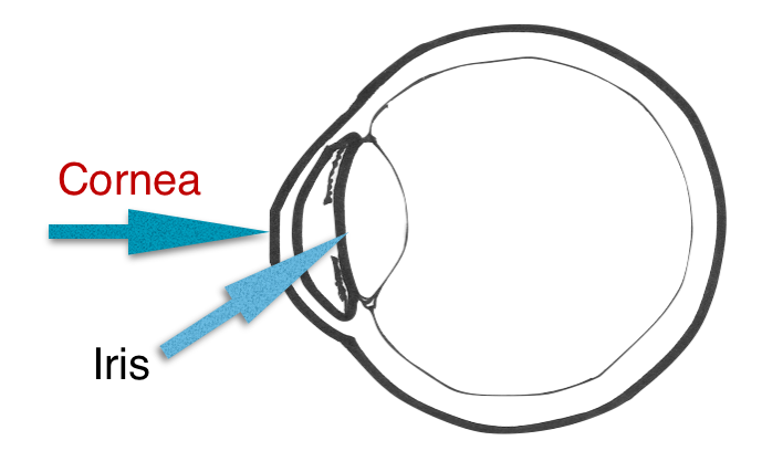 The cornea is the very front part of the eye. It's the clear, circular dome shaped outer most part of the eye. The colored part, or iris, is flat right underneath it.
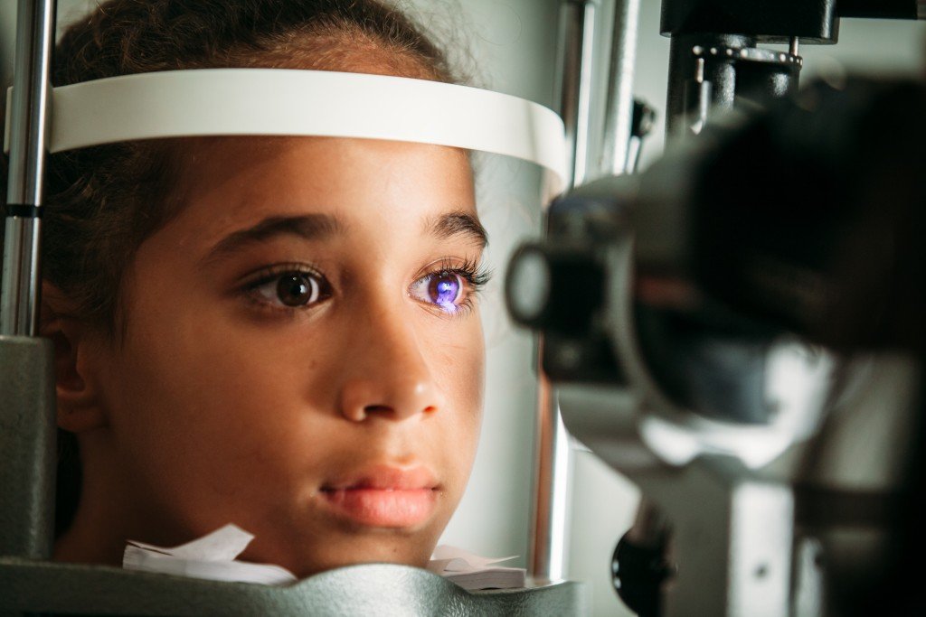 Now, lots of people have astigmatism. Astigmatism is when the cornea is not perfectly round. I usually tell patients and their parents int he exam room, that most corneas look spherical, like a basketball.
  Corneas with astigmatism or more egg or football shaped.
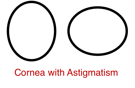
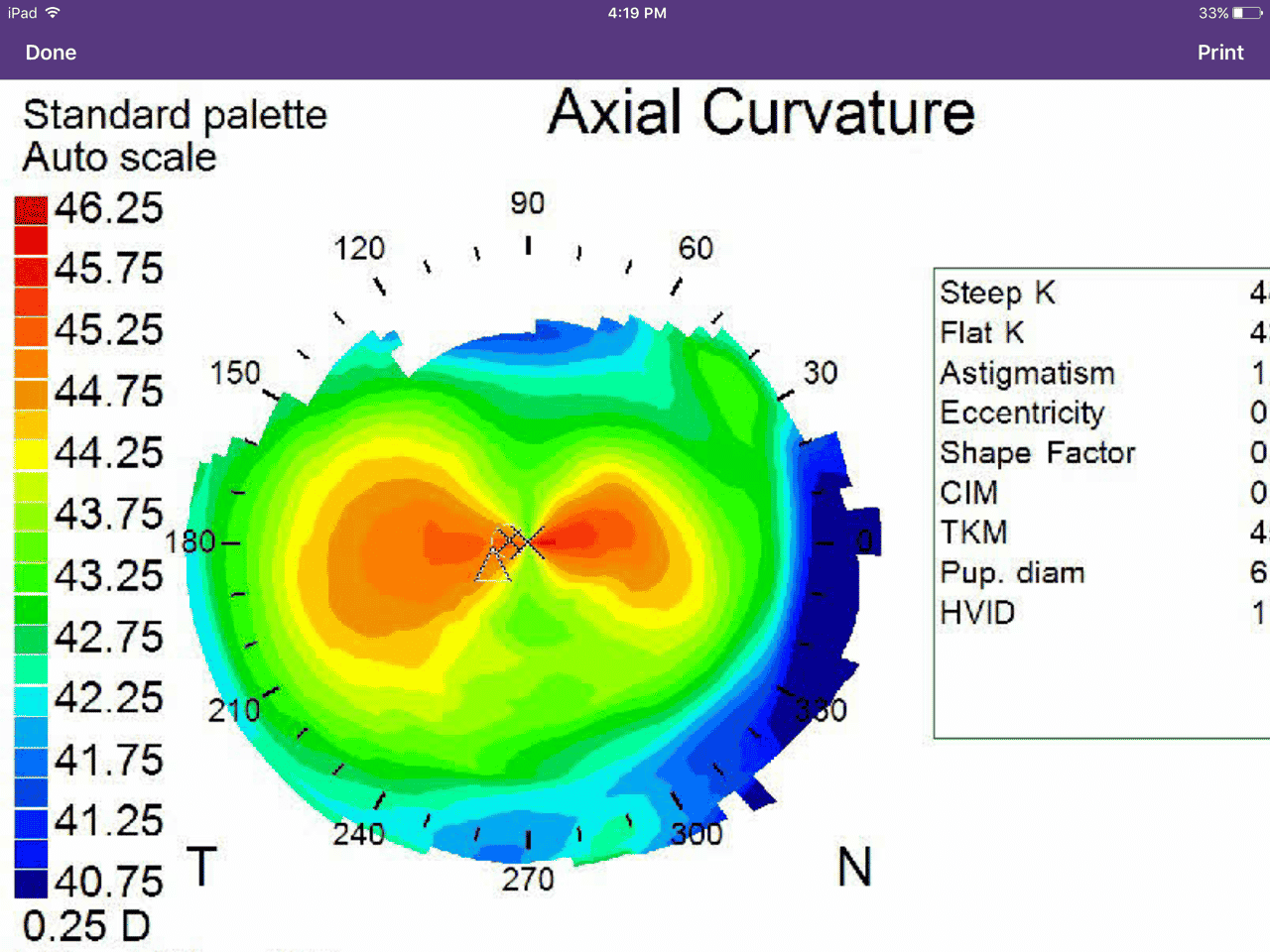
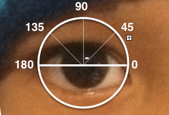 One axis of the circle is a little longer than the other.But, people with keratoconus, don't just have regular astigmatism. They have irregular astigmatism. It's common, occurring 50 people per 100,000. And, unlike regular astigmatism, which usually stays pretty stable throughout life, keratoconus is progressive. The center part of the cornea starts to become thinner and bulges. This usually worsens during puberty, which is why many people are first diagnosed in their teens.Since their corneas are thin, patients shouldn't be playing contact sports. One of the hardest things I had to do was tell a 15 year old, up and coming MMA fighter that he couldn't fight anymore.This is a topography, or map of the cornea in a normal eye. There's a nice symmetric, red bow-tie in the middle. This patient has regular astigmatism.
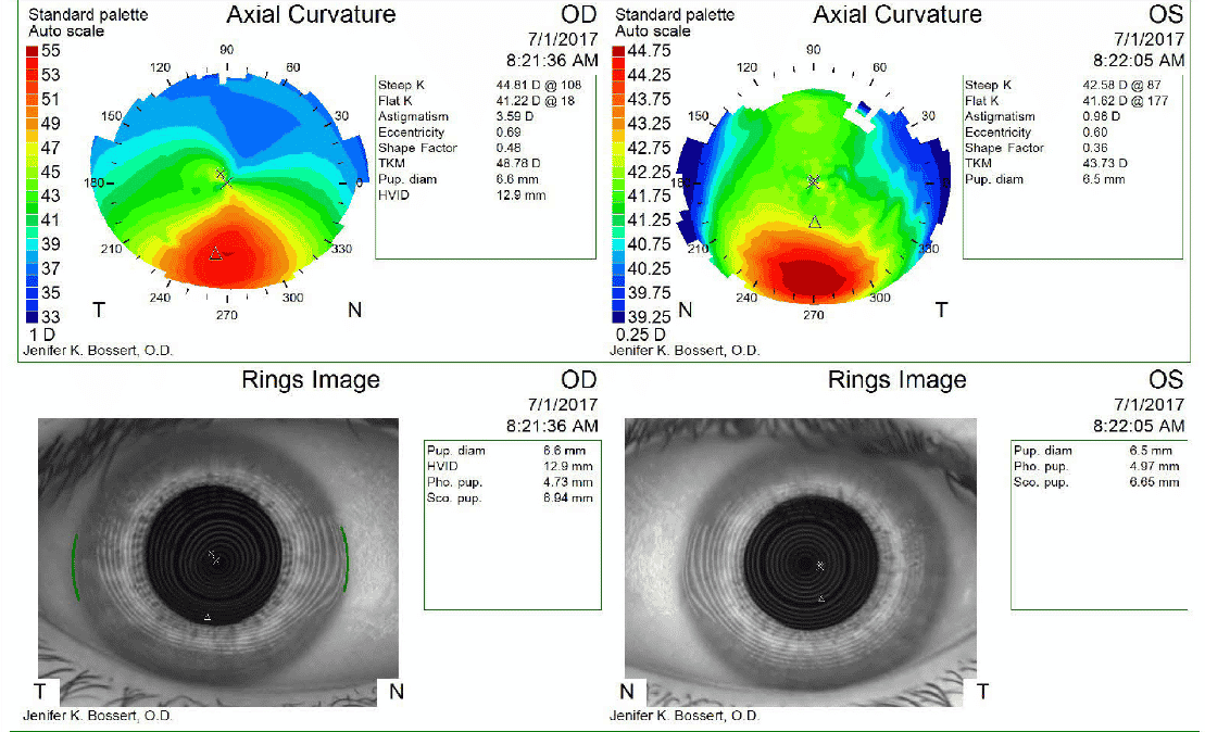
 And, here's a kid with keratoconus. See how it has circles of red at the bottom of both eyes? That means the cornea is really steep there.
 In the past, we would just advise teenage patients to stop all contact-sports and offer contact lenses to improve their vision. But, now, there is a new FDA approved treatment to stabilize progressive keratoconus. It consists us using UV light to stiffen the collagen in the cornea, along with riboflavin eye drops.
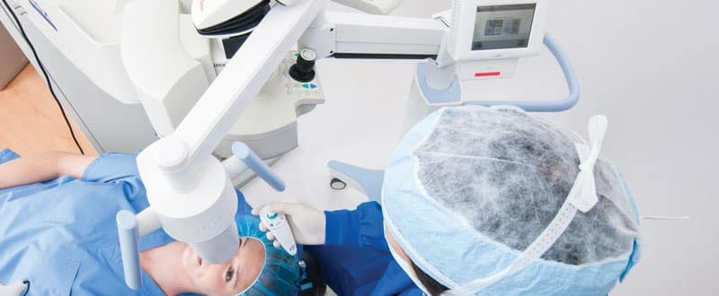

 Drawing of Collagen Before Cross Linking Collagen After Cross Linking (stronger and thicker)The patient above actually traveled from Hawaii to Seattle to get the treatment, but now, one of the Cornea doctors on the island can perform this procedures. It is ideal in younger patients, since it does not reverse damage that has already occurred. Unfortunately, it's not a cure. But, it stabilizes their keratoconus thinning, so if we catch kids early enough, then we can offer treatment which can truly change their ocular health. Of course, all surgery has risks, and this article nicely discusses them, but I think it's wonderful that it's finally FDA approved in this country. We're hoping that insurance companies will realize this procedure prevents potential future blindness and disability and start covering it.Severe cases of keratoconus require treatment with a corneal transplant or INTACS, which I will save for a later post.Two of the kids who I saw in the office were initially misdiagnosed as having "amblyopia". Amblyopia simply means poor vision in one eye. This can be typically caused by an unequal refractive error between two eyes, misaligned eyes, or strabismus, and something obstructing vision in the eye. There should be always be a REASON for the poor vision. And, in these two children's cases, there was a reason - keratoconus. One patient was only 9 years old and the other 13 year old, which is definitely on the young side. But, the fantastic thing is that now there is something that can be done to prevent keratoconus from getting worse.

